Dominio hemisférico del lenguaje analizado con resonancia magnética DTI: correlación con el test de Wada
Dentro de la línea de investigación clínica, el equipo del Dr. García de Sola ha publicado recientemente otro trabajo sobre la lateralización del lenguaje en algunos pacientes con epilepsia farmacorresistente.
Dominio hemisférico del lenguaje analizado con resonancia magnética DTI: correlación con el test de Wada
OBJETIVO
La lateralización del lenguaje es una preocupación importante en algunos pacientes con epilepsia farmacorresistente que se someterán a una cirugía; en estos pacientes, la prueba de dominancia hemisférica es esencial para evitar mayores complicaciones. La prueba de Wada se considera el examen de referencia para la localización del lenguaje, pero es invasiva y requiere muchos recursos humanos y materiales. La resonancia magnética funcional y la tractografía con imágenes con tensor de difusión (DTI) han demostrado que podrían ser útiles para la localización del lenguaje en la cirugía de la epilepsia, pero no hay pruebas de la correlación entre la prueba de Wada y la resonancia magnética con DTI en la dominación del lenguaje.
MÉTODOS
Los autores realizaron una revisión retrospectiva de los pacientes que se sometieron a la prueba de Wada antes de la cirugía de epilepsia en su institución de 2012 a 2017. Los autores analizaron retrospectivamente la anisotropía fraccionada (FA), el número y la longitud de las fibras y el volumen del fascículo arqueado y del fascículo no coordinado, comparando los hemisferios dominantes y no dominantes.
RESULTADOS
Se revisaron diez pacientes con epilepsia del lóbulo temporal. El análisis estadístico mostró que la FA media del fascículo arqueado en el hemisferio dominante era más alta que en el hemisferio no dominante (0,369 vs 0,329, p= 0.049). Además, el número de fibras en el arcado fasciculus era mayor en el hemisferio dominante (881,5 vs 305,4, p = 0,003). Sin embargo, no se encontraron diferencias en el AF del fascículo no cincado o en el número de fibras entre los hemisferios. La longitud de las fibras del fascículo no compactado era más larga en el lado dominante (74,4 vs 50,1 mm, p = 0,05). El volumen en ambos haces era más prominente en el hemisferio dominante (12,12 vs 6,48 cm3, p = 0,004, en el fascículo arqueado, y 8,41 vs 4,16 cm3, p = 0,018, en el fascículo no cerrado). Finalmente, estos parámetros se compararon en pacientes en los que el foco de la convulsión estaba situado en el hemisferio dominante: FA (0,37 vs 0,30, p = 0,05), número de fibras (114,4 vs 315,6, p = 0,014), y el volumen (12,58 vs 5,88 cm3, p = 0,035) en el fascículo arqueado se encontró que era estadísticamente significativo en los hemisferios dominantes. El análisis discriminante lineal del AF, el número de fibras y el volumen del fascículo arqueado mostró una discriminación correcta en el 80% de los pacientes (p = 0,024).
CONCLUSIONES
El análisis del fascículo arqueado y otros haces de tracto por DTI podría ser una herramienta útil para la prueba de localización del lenguaje en el estudio preoperatorio de pacientes con epilepsia refractaria.
PALABRAS CLAVE
Prueba de Wada; MRI; DTI; imagen de tensor de difusión; fascículo arqueado; fascículo incinerado; anisotropía fraccionada; dominación del hemisferio; epilepsia
Language hemispheric dominance analyzed with magnetic resonance DTI: correlation with the Wada test
Juan Delgado-Fernández, MD,1 Maria Ángeles García-Pallero, MD,2
Rafael Manzanares-Soler, MD,3 Pilar Martín-Plasencia, PhD,4 Guillermo Blasco, MD,4 Natalia Frade-Porto, MD,4 Marta Navas-García, MD,4 Paloma Pulido, PhD,4 Rafael G. Sola, PhD,5,6 and Cristina V. Torres, PhD4
1 Department of Neurosurgery, University Hospital 12 de Octubre, Madrid;
2 Department of Neurosurgery, Hospital Universitario Central de Asturias, Asturias;
3 Department of Radiology and
4 Department of Neurosurgery, University Hospital La Princesa, Madrid;
5 Department of Innovation in Neurosurgery, Universidad Autonoma de Madrid; and
6 Department of Neurosurgery, Hospital del Rosario, Madrid, Spain
Language hemispheric dominance analyzed with magnetic resonance DTI: correlation with the Wada test
OBJECTIVE. Language lateralization is a major concern in some patients with pharmacoresistant epilepsy who will face surgery; in these patients, hemispheric dominance testing is essential to avoid further complications. The Wada test is considered the gold standard examination for language localization, but is invasive and requires many human and material resources. Functional MRI and tractography with diffusion tensor imaging (DTI) have demonstrated that they could be useful for locating language in epilepsy surgery, but there is no evidence of the correlation between the Wada test and DTI MRI in language dominance.
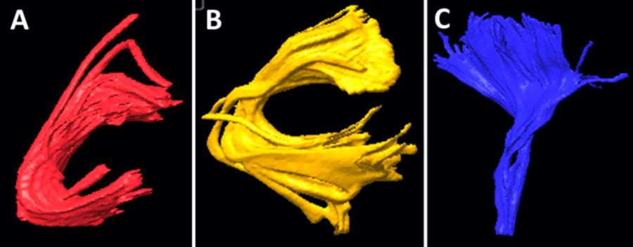
METHODS. The authors performed a retrospective review of patients who underwent a Wada test before epilepsy surgery at their institution from 2012 to 2017. The authors retrospectively analyzed fractional anisotropy (FA), number and length of fibers, and volume of the arcuate fasciculus and uncinate fasciculus, comparing dominant and nondominant hemispheres.
RESULTS. Ten patients with temporal lobe epilepsy were reviewed. Statistical analysis showed that the mean FA of the arcuate fasciculus in the dominant hemisphere was higher than in the nondominant hemisphere (0.369 vs 0.329, p = 0.049). Also, the number of fibers in the arcuate fasciculus was greater in the dominant hemisphere (881.5 vs 305.4, p = 0.003). However, no differences were found in the FA of the uncinate fasciculus or number of fibers between hemispheres. The length of fibers of the uncinate fasciculus was longer in the dominant side (74.4 vs 50.1 mm, p = 0.05). Volume in both bundles was more prominent in the dominant hemisphere (12.12 vs 6.48 cm3, p = 0.004, in the arcuate fasciculus, and 8.41 vs 4.16 cm3, p = 0.018, in the uncinate fasciculus). Finally, these parameters were compared in patients in whom the seizure focus was situated in the dominant hemisphere: FA (0.37 vs 0.30, p = 0.05), number of fibers (114.4 vs 315.6, p = 0.014), and volume (12.58 vs 5.88 cm3, p = 0.035) in the arcuate fasciculus were found to be statistically significantly higher in the dominant hemispheres. Linear discriminant analysis of FA, number of fibers, and volume of the arcuate fasciculus showed a correct discrimination in 80% of patients (p = 0.024).
CONCLUSIONS. The analysis of the arcuate fasciculus and other tract bundles by DTI could be a useful tool for language location testing in the preoperative study of patients with refractory epilepsy.
KEYWORDS: Wada test; MRI; DTI; diffusion tensor imaging; arcuate fasciculus; uncinate fasciculus; fractional anisotropy; hemisphere dominance; epilepsy
Background
Language is one of the most complex functions of the brain and involves different cortical areas and white matter fascicules. Classically, the theory of language based on the anatomical findings of Wernicke
and Geschwind described the interconnection of the Broca and Wernicke areas by the arcuate fasciculus. Currently, the dual-stream theory of language includes the dorsal stream, involved in sensorimotor features of the language, and the ventral stream for speech comprenhension.1 This model also includes the uncinate fasciculus (among others), which connects the inferior longitudinal fasciculus to the inferior frontal area and is one of the main tracts on the ventral stream, connecting both pathways.1,2
Language is a lateralized function in most humans: 95% of right-handed people show left-sided hemispheric localization, while 15% of left-handed people show rightsided localization.3 However, in some cases an atypical language lateralization can be identified. Different studies have demonstrated that in patients with previous lesions in the left hemisphere, language lateralization is influenced.4Ð7 This is the case in epilepsy patients who have demonstrated a high degree of language circuit reorganization, mainly in intractable seizures.8,9
In this context, a precise location of the epileptogenic focus is mandatory, as surgery should be tailored to avoid damaging any underlying structures.10 Normally, the epilepsy protocol includes scalp EEG, neuropsychological evaluation, MRI, and PET or SPECT,11,12 but in other cases it should be extended with stereoEEG and foramen ovale electrodes, or subdural electrodes.13 Nevertheless, some patients without good outcomes in epilepsy surgery could benefit from further imaging, such as diffusion tensor imaging (DTI).14,15
DTI has demonstrated its utility in describing the microstructure of white matter tracts and defining fascicules and bundles in preoperative planning. In addition, fractional anisotropy (FA), which describes the degree of anisotropy of a diffusion process,16 is a very useful tool that helps us to understand the structural integrity of axons and myelin sheaths.17,18
Moreover, there are a large number of studies that have recently demonstrated a correlation between DTI and functional MRI (fMRI), which could help us define language lateralization through DTI, thereby avoiding invasive tests such as the Wada test.8,9,19Ð22 However, the Wada test still remains the gold standard test to determine language lateralization, and if we assume that DTI correlates with Wada test results, that could lead to a possible bias. Moreover, the Wada test and fMRI do not have a perfect correlation. In a meta-analysis, Dym et al.23 reviewed 442 patients, comparing fMRI with the Wada test, and showed that sensitivity and specificity of fMRI for atypical language dominance were 83.5% and 88.1%, respectively.
Nevertheless, although the Wada test remains the only definitive method for defining language dominance, it is an invasive, costly, and unpleasant experience for patients.24 Therefore, the objective of our study was to compare our results from a preoperative Wada test to DTI, so DTI could be used for preoperative language lateralization in the future.
…
Conclusions
Recent studies have stated that DTI could help determine language dominance in patients. However, we believe it is very important to remember that, before we can assume that DTI could replace an invasive technique such as the Wada test, we have to get a better correlation between DTI and the gold standard test for language location, meaning that DTI should be compared with the Wada test, and not compare it with fMRI. This topic has been little studied and more investigation should be performed, but some previous investigations and the present study make some favorable arguments that the dominant hemisphere could be detected, not only through the presence of higher DTI parameter values such as FA, volume, and number and length of fibers (mainly in the arcuate fasciculus), but also in other fiber bundles such as the uncinate fasciculus, and other tracts involved in language as technology evolves.